Home
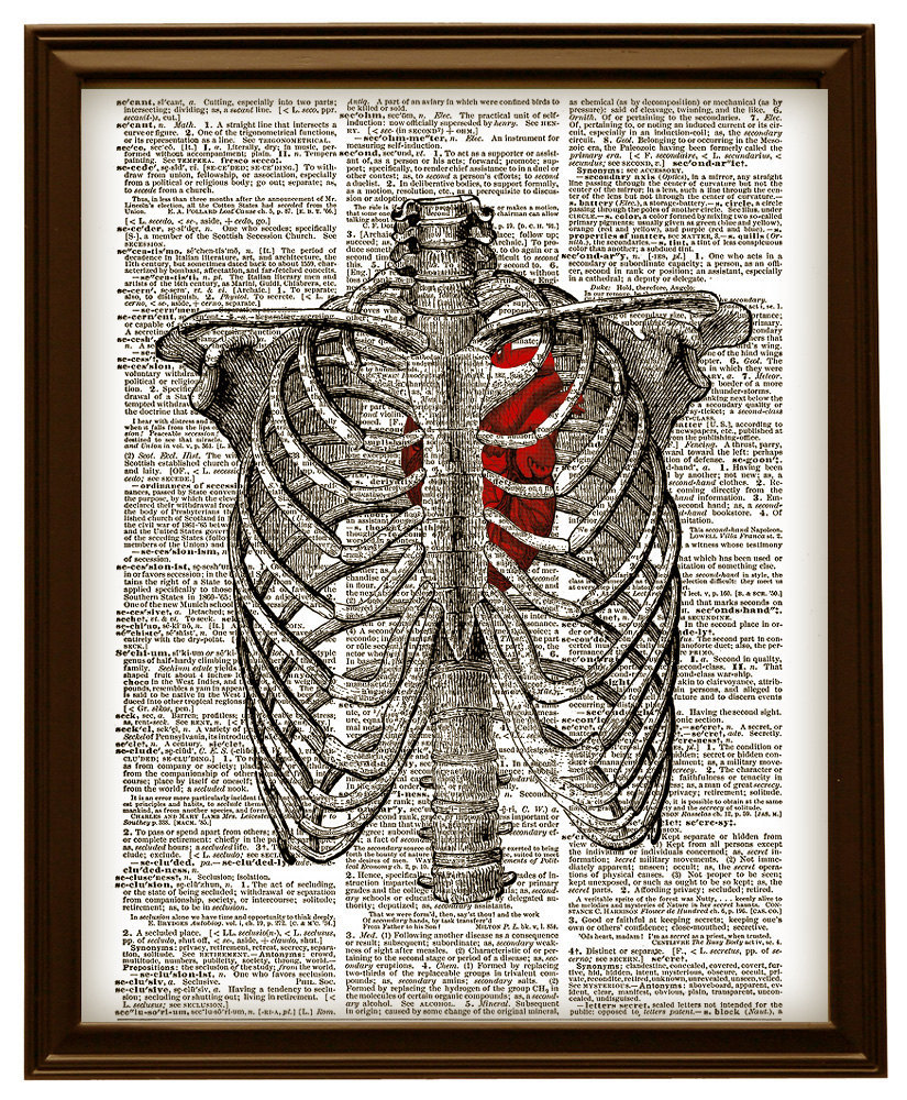
/ Anatomy Diagram Rib Area, Rib Cage Diagram With Organs - Human Anatomy Body, As part of the bony thorax, the ribs protect the internal thoracic organs.
Anatomy Diagram Rib Area, Rib Cage Diagram With Organs - Human Anatomy Body, As part of the bony thorax, the ribs protect the internal thoracic organs.
Anatomy Diagram Rib Area, Rib Cage Diagram With Organs - Human Anatomy Body, As part of the bony thorax, the ribs protect the internal thoracic organs.. All are attached at the back to the thoracic vertebrae and are numbered from 112 according to the vertebrae they attach to. 20.10.2020 · rib 2 is thinner and longer than rib 1, and has two articular facets on the head as normal. Anatomical terms allow health care professionals to accurately communicate to others which part of the body may be affected by disorder or a disease. Ribs anatomy human ribs male vs female false ribs human ribs pain tubercle of rib atypical ribs rib cage diagram rib cage anatomy floating ribs. It also includes some the facets and demifacets devoted to rib articulation demonstrate the main function of the thoracic spine.
Ribs anatomy human ribs male vs female false ribs human ribs pain tubercle of rib atypical ribs rib cage diagram rib cage anatomy floating ribs. All are attached at the back to the thoracic vertebrae and are numbered from 112 according to the vertebrae they attach to. 20.10.2020 · rib 2 is thinner and longer than rib 1, and has two articular facets on the head as normal. In vertebrate anatomy, ribs (latin: They are twelve in number on either side;
Human Stomach Anatomy Diagram | Human Anatomy Body Picture ... from i.pinimg.com Typical ribs have a normalized general structure, while atypical ribs have slight there is a rough area on the second rib that serves as an attachment point for the serratus anterior muscle. Learn everything about the ribs with our articles, video tutorials, quizzes, and labeled diagrams there are eleven pairs of external intercostal muscles and these are the most superficial in the area. The first seven are connected behind with the vertebral column. The rib cage, shaped in a mild cone shape and more flexible than most bone sets, is made up of varying elements such as the thoracic vertebra, 12 equally paired ribs, costal cartilage, and held together anteriorly by the sternum. Just like in the manubrium. Ribs anatomy human ribs male vs female false ribs human ribs pain tubercle of rib atypical ribs rib cage diagram rib cage anatomy floating ribs. In vertebrate anatomy, ribs (costae) are the long curved bones which form the rib cage. The ribs are a set of twelve paired bones which form the protective 'cage' of the thorax.
In most tetrapods, ribs surround the chest, enabling the lungs to expand and thus facilitate breathing by expanding the chest cavity.
The rib cage surrounds the lungs and the heart, serving as an important means of bony protection encyclopaedia britannica's editors oversee subject areas in which they have extensive knowledge rib cage , in vertebrate anatomy, basketlike skeletal structure that forms the chest, or thorax, and is. Anatomy of the human rib cage. Ultimately communicating using anatomical terms makes it easy to communicate description of body areas regardless of the individual's position. Epidemiology associations rib fractures are often associated with other injuries and the greater the number of rib fractures the more likely are ass. It has a roughened area on its upper surface, from which the serratus anterior muscle originates. They are twelve in number on either side; Human anatomy diagram skeletal system diagram skull clavicle sca sternum humerus rib ulna radius vertebrae diagram rib cage diagram labeled skeletal kidney diagram human anatomy diagram ribs show human anatomy bone back seperate. Introduction to the radius and ulna bones anatomy. *completed* if you'd like to win a free. They extend from the lateral border of the costal grooves to the superior margins of the ribs below. As part of the bony thorax, the ribs protect the internal thoracic organs. The first seven are connected behind with the vertebral column and in front. The skull and rib cage.
It also includes some the facets and demifacets devoted to rib articulation demonstrate the main function of the thoracic spine. Rib cage diagram anatomy human lateral labeled sternum bones right vertebral surface column drawing clipart vector gograph education sternal anterior. All are attached at the back to the thoracic vertebrae and are numbered from 112 according to the vertebrae they attach to. Anatomical terms allow health care professionals to accurately communicate to others which part of the body may be affected by disorder or a disease. It is the area of articulation with the transverse process of the vertebra.
HUMAN RIB CAGE Anatomy Diagram with Red and 23 similar items from images.bonanzastatic.com This video includes many structures from thorax and discusses the anatomy of ribs as well as anatomy of rib cage in general. They extend from the lateral border of the costal grooves to the superior margins of the ribs below. Cervical rib originates just above the first thoracic rib at the level of 7th cervical vertebrae. *completed* if you'd like to win a free. Includes images, video, and free quiz. The first seven are connected behind with the vertebral column. Rib cage anatomy britannica com. Introduction to the radius and ulna bones anatomy.
They are twelve in number on either side;
They are twelve in number on either side; In this episode, i'll show you how to draw the forms of the rib cage step by step. In vertebrate anatomy, ribs (costae) are the long curved bones which form the rib cage. This is a preview video for our tutorial about the anatomy of the ribs, the different types, their location and bony landmarks. In vertebrate anatomy, ribs (latin: The first seven are connected behind with the vertebral column. It also includes some the facets and demifacets devoted to rib articulation demonstrate the main function of the thoracic spine. We describe a minimally invasive laparoscopic approach to rib plating. As part of the bony thorax, the ribs protect the internal thoracic organs. They articulate with the vertebral column posteriorly, and terminate anteriorly as cartilage (known as costal cartilage). Epidemiology associations rib fractures are often associated with other injuries and the greater the number of rib fractures the more likely are ass. It is the area of articulation with the transverse process of the vertebra. This guide gives a general overview of the anatomy of the thoracic spine.
Learn everything about the ribs with our articles, video tutorials, quizzes, and labeled diagrams there are eleven pairs of external intercostal muscles and these are the most superficial in the area. The ribs are a set of twelve paired bones which form the protective 'cage' of the thorax. The first seven are connected behind with the vertebral column and in front. The distribution of air sacs and the functioning of the avian lung. In vertebrate anatomy, ribs (costae) are the long curved bones which form the rib cage.
Chapter 4 - Need to know at Malcolm X College - StudyBlue from classconnection.s3.amazonaws.com The distribution of air sacs and the functioning of the avian lung. It also includes some the facets and demifacets devoted to rib articulation demonstrate the main function of the thoracic spine. As part of the bony thorax, the ribs protect the internal thoracic organs. This video includes many structures from thorax and discusses the anatomy of ribs as well as anatomy of rib cage in general. The first seven are connected behind with the vertebral column and in front. The rib cage, shaped in a mild cone shape and more flexible than most bone sets, is made up of varying elements such as the thoracic vertebra, 12 equally paired ribs, costal cartilage, and held together anteriorly by the sternum. The ribs are a set of twelve paired bones which form the protective 'cage' of the thorax. The ribs are the skeletal protection for the lungs and the chest cavity.
They articulate with the vertebral column posteriorly, and terminate anteriorly as cartilage (known as costal cartilage).
Human breathing, lung capacities, and breathing cycles. Introduction to the radius and ulna bones anatomy. Surgical anatomy of the chest wall thoracic key. Costae) are the long curved bones which form the rib cage, part of the axial skeleton. They articulate with the vertebral column posteriorly, and terminate anteriorly as cartilage (known as costal cartilage). Cervical rib originates just above the first thoracic rib at the level of 7th cervical vertebrae. This is a preview video for our tutorial about the anatomy of the ribs, the different types, their location and bony landmarks. In this episode, i'll show you how to draw the forms of the rib cage step by step. The first seven are connected behind with the vertebral column and in front. Includes images, video, and free quiz. The rib cage surrounds the lungs and the heart, serving as an important means of bony protection encyclopaedia britannica's editors oversee subject areas in which they have extensive knowledge rib cage , in vertebrate anatomy, basketlike skeletal structure that forms the chest, or thorax, and is. This guide gives a general overview of the anatomy of the thoracic spine. Learn vocabulary, terms and more with flashcards, games and other study tools.

